Ultrasound Image Quality
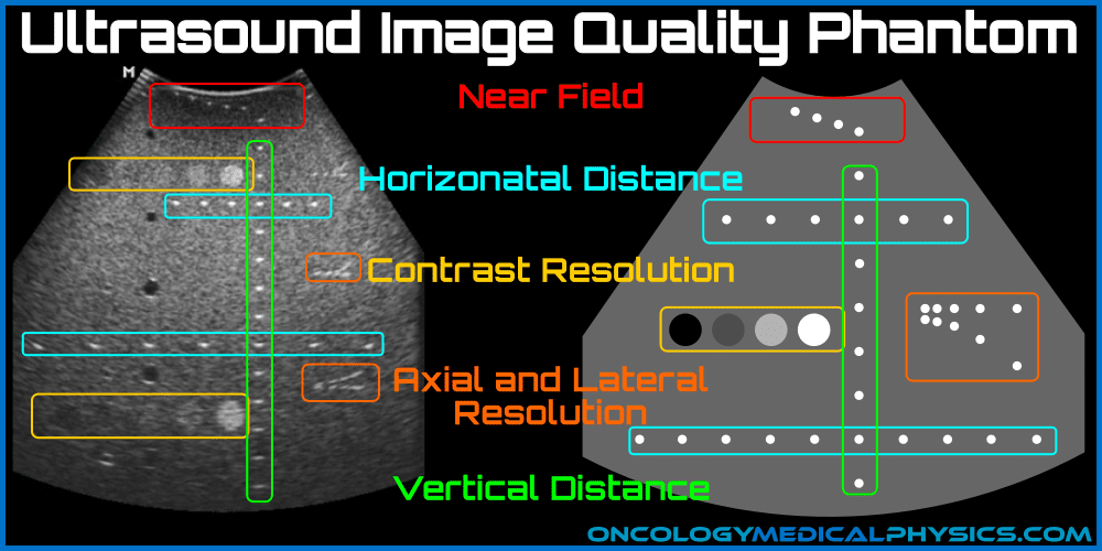
Image Quality Phantom
A phantom is a device used to asses the quality of ultrasound images. Most ultrasound phantoms consist of a water like substance with high and low density objects embedded at known locations.
Spatial Resolution
Axial Resolution
Axial resolution refers to the ability to distinguish closely spaced objects along the direction of beam propagation. The minimal object spacing that can be resolved is ½ spatial pulse length (SPL). SPL is determined by the number of pulses emitted per cycle, the wavelength, and ring down time.
Typical Axial resolution = 0.5 mm for a 5 MHz beam.
Highest axial resolution is achieved with:
- Low Q-factor (heavy damping)
- Low wavelength/high frequency
Lateral Resolution
Lateral spatial resolution, sometimes called azimuthal resolution, is the ability to discern two closely spaced objects oriented perpendicular to the beam. As the beam converges and then diverges, lateral spatial resolution is dependent upon the distance to the object. Lateral spatial resolution of linear and curvilinear arrays can be varied by changing the number of elements firing as a group.
Highest lateral resolution is achieved with:
- Objects located at the end of the near field
- Small groups of elements firing
Slice Thickness
Slice thickness, sometimes called elevational resolution, is the width of the imaging plane. Slice thickness depends on the height of the transducer element and is usually the lowest resolution axis. This low spatial resolution leads to partial volume averaging, in which the intensity of a pixel is the average of all reflection intensities within a slice.
Some arrays, called multi-linear arrays, have multiple rows of elements which allow the transducer to adjust slice thickness by adjusting the number of rows firing.
Contrast Resolution
Contrast resolution refers to the ability to discern objects of differing intensities. This is dependent upon the strength of reflection (returning signal) and the image noise. The concepts of return signal and noise are often combined into a single value called the signal-to-noise ratio (SNR).
Contrast resolution is highest for:
- Variables which maximize the strength of an acoustic echo:
- Objects with very different acoustic impedance to their surroundings
- Variables which reduce partial volume averaging:
- Higher spatial resolution
- Higher temporal resolution - minimizing impact of motion
- Variables which maximize signal-to-noise ratio:
- Use of higher ultrasound power requiring less electronic signal amplification
- Superficial objects subject to less attenuation and returning scatter
Key point: The Rose Criteria states that an SNR of 5 or more is needed to identify image features with certainty. An SNR of less than 5 indicates that there will be difficulty and uncertainty in identifying low contrast image features.
Temporal Resolution (Frame rate)
The temporal resolution, also referred to as frame rate, of a 2D image is the time required to acquire all A-lines used in the B-mode image.

- c is the speed of sound in tissue
- N(A-lines) are the number of A-lines required to produce a B-mode 2D image
- D is the depth of scan; 2D is used because the ultrasound wave must travel to the depth of scan and back to the transducer
Highest temporal resolution is achieved with:
- A low number of A-lines
- A low depth of scan
Navigation
Not a Premium Member?
Sign up today to get access to hundreds of ABR style practice questions.


