
Statistical Radiobiology
Linear Quadratic Model (LQM)
The linear quadratic model is the most commonly used model of cell survival used in radiation therapy. LQM is clinically useful in determining the efficacy of a given fractionation scheme.
![]()
- D is the dose administered
- S(D) is the fraction of cells to survive a given dose.
- αD is the probability of cell death arising from a single "double hit" producing a double strand break.
- βD2 is the probability of cell death arising from multiple "single hits," each generating single strand breaks, close enough together to cause a double strand break.
LQM Assumptions
- DNA hits, events that damage DNA, are random with probability proportional to dose.
- Double strand breaks are required for cell sterilization (i.e. death or, equivalently, inability to reproduce)
- There are two methods of producing a double strand break
- One quanta of radiation damages both DNA strands (αD). This is referred to as a "double hit" because it damages two strands with one hit.
- Two quanta of radiation each breaking a single strand, produce a double strand break (βD2). These are referred to as "single hits" because one strand is damaged with each hit.

Alpha/Beta Ratio
Alpha/Beta ratio relates the relative importance of single and double strand breaks in causing cell death. In effect, alpha/beta ratio indicates how resistant a cell is to radiation damage.
High alpha/beta ratios (around 10) indicate that single hit damage does not readily accumulate to lethal effects and there is little increase in cell killing per unit dose for higher total doses. That is, high alpha/beta ratios indicate a linear plot on the cell survival plot.
Low alpha/beta ratios (1-3) indicate that the accumulation of multiple single hits produces increased lethality for higher doses. Cell survival plots for low alpha/beta ratio cells have a greater curvature.
Data from Technical Basis of Radiation Therapy, 5th Edition Table 1
| Tissue Type | Alpha/Beta |
|---|---|
| Tumor and Early Effects | 10 |
| Late Complications | 3 |
| Late CNS Effects | 2 |
| Prostate | 1.5 |

Key Point: High alpha/beta ratio tumors benefit from fractionation because it reduces normal tissue toxicity without significant reduction in tumor lethality.
Fractionation
Fractionation is the division of a treatment dose into several discrete treatments (fractions). Fractionation allows the oncology team to leverage the differences in alpha/beta ratio between tumors (~10) and normal tissue (~3) to improve the therapeutic ratio.
Common Fractionation Schemes
Standard Fractionation
- 1.8 - 2Gy per fraction
- 1 treatment per day, Monday - Friday
- Assuming 109 tumor cells and an expected kill ratio of 50% per 2Gy fraction, 30 fractions is sufficient to reduce the number of expected surviving cells to less than 1.
BID (Hyperfractionation)
- 2 fractions delivered per day (separated by at least 6 hours)
- 1.2Gy per fraction
- Reduced late effects such as those to the central nervous system
- Early effects, such as those to the skin or GI tract, are unchanged
HypoFractionation
- >2Gy per fraction
- Up to 1 fraction per day
- Increases late effected
- Decreases early effects
Radiosurgery
- 1-5 fractions with doses ranging between 8 and 90 Gy per fraction
- Such high dose fractions potentially invalidate the linear quadratic model and are currently not well understood.
- Radiosurgery focuses on avoiding dose to normal tissue rather than on improving therapeutic ratio. As a result, it is commonly used only for small lesions and special cases such as trigeminal neuralgia.
Factors in Fractionated Radiotherapy (The Five Rs)
1. Repair
Sublethal damage is repaired in both tumors and healthy cells. Differences in repair rate may be exploited.
2. Repopulation
Cell division and population growth occurs, albeit to an inhibited degree, between fractions.
3. Reoxygenation
Tumors often have poor vasculature and, as a result, are anoxic. This lack of oxygen makes tumor cells more radioresistant. Fractionation allows time for some tumor cells to die which improves oxygenation of the remaining cells. This effect increases radiosensitivity during subsequent fractions.
4. Redistributions
The distribution of cells in a given cell cycle stage changes with fractionation.
5. Radiosensitivity
Cells differ in their intrinsic radiosensitivity. Radiosensitive cells include haematological cells and epithelial cells as well as haematological tumor cells. Radioresistant cells include neurons and myocytes as well as melanoma and sarcoma tumor cells.
Key Point: 100% kill is not required for long-term survival without recurrence. Rather, it may be sufficient to eradicate the metastatic spread and bring the tumor into partial remission.
Evaluating Fractionation Schemes
Biologically Effective Dose (BED)
Biologically effective dose allows for simple assessment of the biological effect of a particular dose and fractionation scheme, given the alpha/beta ratio of the tissue in question. Two fractionation schemes are equally effective when their BED values are equal.
![]()
- n is the number of fractions delivered
- d is the dose per fraction
- α/β is the alpha over beta value derived from the linear quadratic model
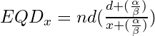
Equivalent Dose (EQD)
Equivalent dose, often notated EQDx where x is the reference dose per fraction, is used to find an equivalent fractionation scheme to a reference scheme. Because standard 2Gy per fraction is most common, EQD2 is most often used.
Key Point: Because of the differences in alpha/beta ratio, an EQD2 cannot be found both for tumor control and normal tissue toxicity simultaneously. Often, the new fractionation scheme will be limited by normal tissue tolerances.
Relative Biological Effectiveness
Relative biological effectiveness (RBE) is the ratio of absorbed dose required to produce an effect under reference conditions to the absorbed dose required to produce the same effect under another set of conditions. RBE is a useful concept because the effect of a given dose is determined not just by the absorbed dose but also by the type of radiation and the circumstances under which the radiation is delivered.
![]()
- Deval is the dose which is being evaluated
- Dref is the reference dose
- NOTE: Doses here are isoeffective dose
Key Point: A positive RBE indicates that the dose under investigation is more effective than the reference dose.
Factors influencing RBE
- Linear Energy Transfer (LET)
- Radiation quality
- Fractionation
- Dose rate
- Biological system in question
- Biological conditions
Oxygen Enhancement Ratio (OER)
Oxygen is a radiosensitizer which improves the RBE compared to anoxic conditions. Oxygen enhancement ratio is a special care of BRE measuring the impact of oxygenation on radiosensitivity.
![]()
- OER for photons, X-rays and gamma rays, is typically between 2.5 and 3
- Neutrons have a low OER, typically around 1.5
Key Point: High LET particles, such as protons and alpha particles, produce more radiation damage per unit path length. This increases the probability of double strand breaks for a given absorbed dose in increases RBE.
Navigation

Not a Member?
Sign up today to get access to hundreds of ABR style practice questions.