Electron Dose Distributions
Electron Depth Dose Distributions
The behavior of electron depth dose distributions is driven by Coulomb interactions with charged subatomic particles. Clinically, this translates into percent depth dose (PDD) distributions with significantly higher surface dose and more rapid dose fall-off than photon beams.
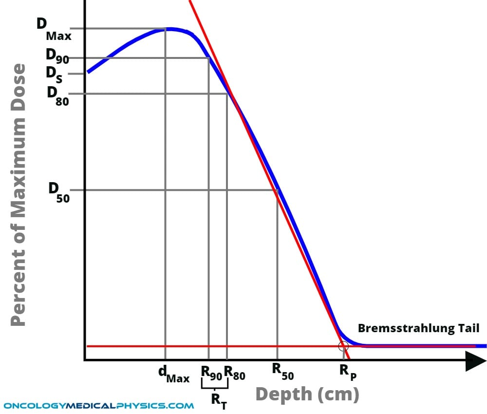
Depth Dose Regions of Interest
- DMax is the maximum dose.
- DS is the surface dose.
- Dx is the dose at x percent of maximum dose.
- dMax is the depth of maximum dose.
- Rx is the depth of x percent of maximum dose.
- RT is the therapeutic range. Therapeutic range is typically taken at either R90 or R80.
- TG-25 and IRCU 35 recommend taking RT as R90.
- RP is the practical range. Practical range is found by extrapolating the linear portion of the depth dose curve and the Bremsstrahlung tail (red lines in the above figure). Rp is found at the intersection of these lines.
- The Bremsstrahlung tail is comprised of photons originating from Bremsstrahlung interactions in the treatment head and the patient/phantom.
- Bremsstrahlung accounts for around 5-15% of dose from electron beam.
- Approximately half of the photons are generated in the treatment head with the other half generated in the phantom.
Approximate electron PDD values
| Energy | Surface Dose | dmax | R90% | R50% |
|---|---|---|---|---|
| 6MeV | 78% | 1.2cm | 1.7cm | 2.3cm |
| 9MeV | 81% | 2.0cm | 2.7cm | 3.5cm |
| 12MeV | 86% | 2.8cm | 3.9cm | 5.0cm |
| 15MeV | 91% | 3.2cm | 4.9cm | 6.3cm |
| 20MeV | 95% | 3.5cm | 6cm | 8.5cm |
Electron Energy
AAPM TG-25: Clinical Electron-Beam Dosimetry (external link) provides guidance on the characterization and determination of electron beam energy.
Energy Notation
![]() is the mean energy incident on the phantom surface.
is the mean energy incident on the phantom surface.
![]()
![]() is the most probably energy at the surface in MeV.
is the most probably energy at the surface in MeV.
![]()
![]() is the mean energy at the depth d.
is the mean energy at the depth d.
![]()
Energy and PDD
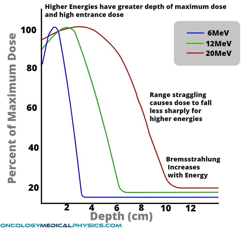
Increased electron energy has the following impacts on a percent depth dose distribution.
- Increases skin dose
- Increases depth of maximum dose
- Increases range straggling
- Decreases sharpness of dose fall-off
- Increases Bremsstrahlung X-ray tail
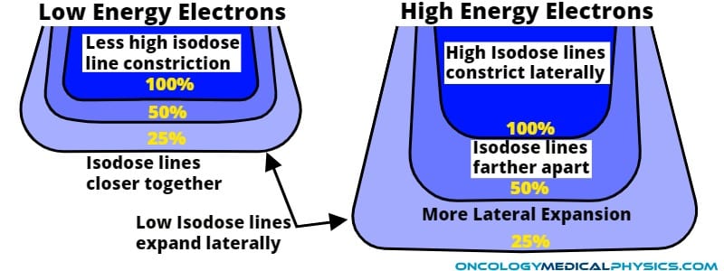
- High dose isodose lines contract slightly
- Low dose isodose lines expand laterally due to range straggling
Approximate Electron Ranges in Water
As a rule of thumb, electrons lose approximately 2MeV per cm in water.
Approximate ranges in water
- R90 (cm) ≅ E0/3.2
- R80 (cm) ≅ E0/2.8
- R50 (cm) ≅ E0/2.33
- Rp (cm) ≅ E0/2
Linear Accelerator Energy Spectrum
The energy spectrum of an electron beam exiting a linac treatment head is an approximately Gaussian distribution centered about an energy just below the accelerating energy. This spectrum results from energy selectors and attenuation straggling in the flattening filters.
The Bremsstrahlung photon component of a clinical energy beam is predominantly low energy and strongly skewed right.
Electron Buildup Explained
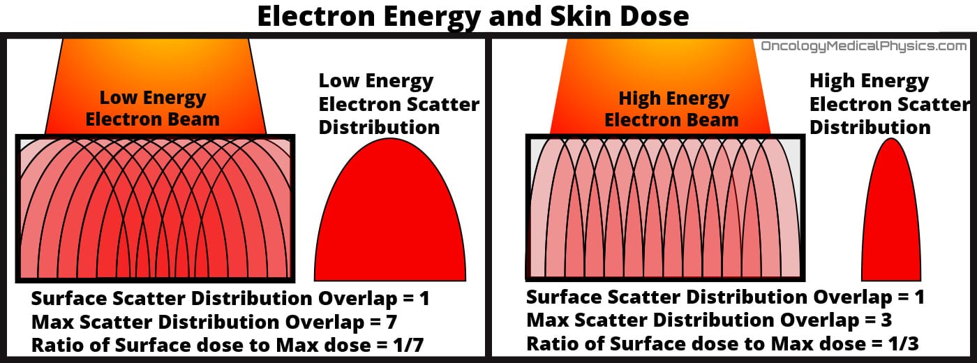
Although stopping power is inversely proportional to the square of velocity, MeV electron velocity does not vary much with energy. As a result, the photon energy loss per unit path length is approximately constant until near the end of the electron's path. How then can we explain the difference in surface dose as an effect of energy?
The answer is found in the impact of electron energy on its scattering angle. Higher energy electrons are scattered in a more forward direction. This means that electrons incident lateral to the point of measurement are less likely to contribute to dose at the point if they are higher energy. The end result is less build-up for higher energy electron beams.
Field Size
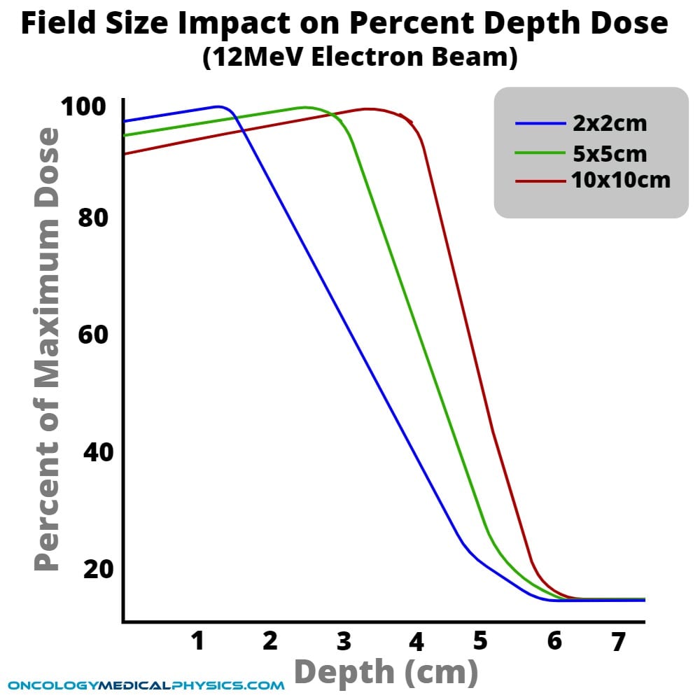
For field sizes smaller than the practical range of incident electrons, lateral charged particle equilibrium is lost and the following trends are observed.
- Decreased field size reduces depth of maximum dose.
- Decreased field size increases relative skin dose.
- Decreased field size increases range straggling and decreases sharpness of dose fall off.
For field sizes larger than the practical range of incident electrons, lateral charged particle equilibrium is preserved and there is little change in depth dose with field size.
Interpolating PDD and field size
PDD may be approximated for a rectangular field size with m (cm) by b (cm) side if the PDD is known for an m by m and b by b field.
![]()
Angle of Incidence
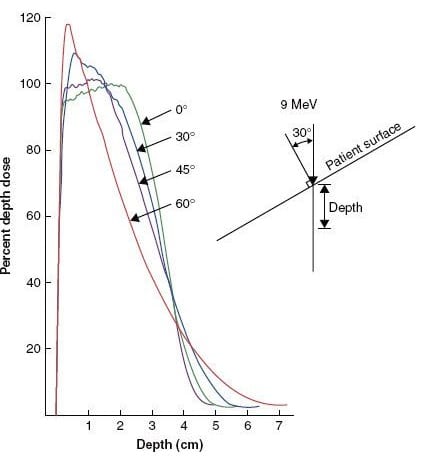
Increased angle of obliquity has the following effects on depth dose distributions:
- Shift the depth of maximum dose, and R80, toward the surface
- Increase Dmax
- Increase surface dose
- Increase the practical electron range
- This is because of the contribution of high scatter angle electrons, which do not pass through as much tissue.
Bremsstrahlung Contamination
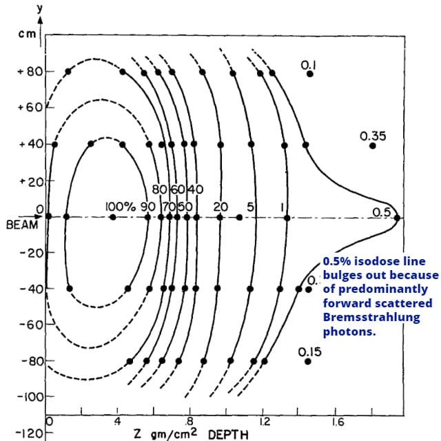
Bremsstrahlung production in clinical electron beams is primarily forward peaked and can result in a bulge along the central axis to the very low isodose lines (0-4%).
Bremsstrahlung contamination increases with
- Increased energy
- Decreased field size
Bremsstrahlung contamination (relative to Dmax)
- <1% at 4Mev
- <2.5% at 10MeV
- <4% at 20MeV
Knowledge Test
This is the sample version of the full quiz. Log in or register to gain access to the full quiz.
Navigation
Not a Member?
Sign up today to get access to hundreds of ABR style practice questions.

