Ocular Brachytherapy
Overview
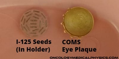
The Collaborative Ocular Melanoma Study (COMS) set the standard for I-125 eye plaque brachytehrapy.
Selected Readings
AAPM TG-129: Dosimetry of 125I and 103Pd COMS Eye Plaques for Intraocular Tumors (external link)
Collaborative Ocular Melanoma Study (COMS) (external link)
Treatment Regimen
Disease: Ocular Melanoma
Prescriptions
- COMS: 85Gy to water at a depth of 5mm (for tumors with apex height of 5mm or less) or to apex depth (if apex is greater than 5mm).
- Max dose is may be as much as 10 times greater than prescription dose (1000Gy)!
- Implant duration: 5-12 days
- Radionuclide: 125I, 103Pd
- Seed activity: 0.5 – 5 mCi
- Dose rate: 0.5 – 1.25 Gy/hr
- Plaque size
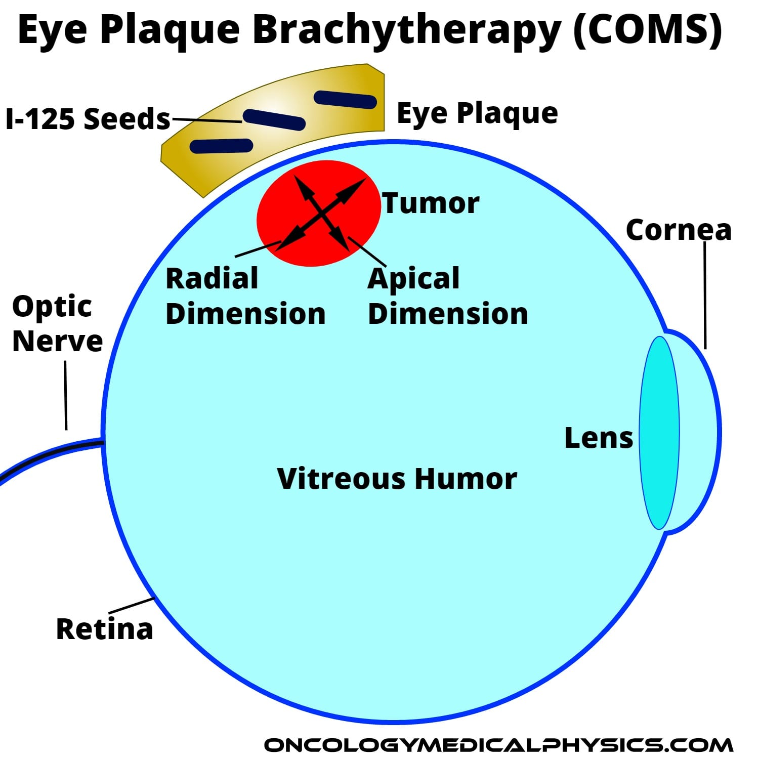
- Chosen to cover radial extent of tumor with 2-3mm margin.
- Available plaque sizes: 10mm to 22mm in 2mm increments
Alternative Treatments
- External beam proton therapy.
- Enucleation (removal of eye).
COMS Planning
- Tumor is localized using Fundoscopy, Ultrasound, CT, or MRI to generate a Fundus diagram.
- Physician determines radial and apical dimensions of tumor using MRI and Fundus diagrams.
- A plaque is chosen exceeding the radial dimensions of the tumor.
- I-125 seeds are ordered in a quantity determined by plaque size and activity determined to yield full dose at tumor apex.
- Sources are assayed upon arrival and entered into inventory.
- Plaque and seeds are assembled and sterilized prior to insertion.
- During the insertion operation, the rectus muscles attaching the eye are cut. The eye is rolled to access tumor and the plaque is stitched in place. The eye is then rolled back to position.
- After 5-12 days the plaque is removed, rector muscles are attached, and patient is released (after survey to verify source removal).
Activity/Dose Calculation
The required activity per seed varies based on the seed design, radionuclide, implant duration, and plaque size. Additionally, some users may asymmetrically load the plaque to yield a tailored dose distribution (e.g. to reduce optic nerve dose). For standard (uniform) seed loading the dose distribution for a reference activity (1Ci) and duration is typically mapped for each plaque size. This may be accomplished either in a treatment planning system, physically using film, or by referencing the table in AAPM TG-129. This dose distribution map can then be by activity to yield the prescription dose at a given depth.
Navigation
Not a Premium Member?
Sign up today to get access to hundreds of ABR style practice questions.


