Contrast and Weighting Schemes
T1 Weighting
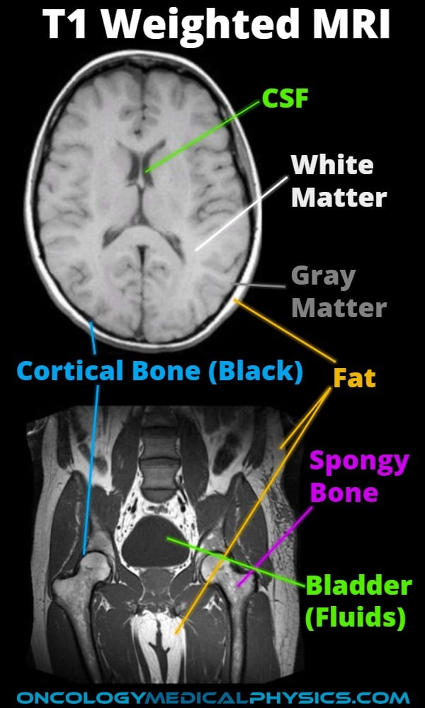
T1 weighting attempts to maximize differences in T1 relaxation while minimizing the impact of T2 relaxation. This is accomplished by using a short TR and TE [i.e. TR approximately equal to T1 (500ms) while TE is much shorter than T2 (<15ms)]. T1 weighting offers good tissue contrast but sees less use in radiation therapy than T2 due to limited tumor contrast. When used in radiation therapy planning, T1 weighting is most often used to asses nodal invasion. The most common T1 weighted sequence is Spoiled Gradient Echo.
T2 Weighting
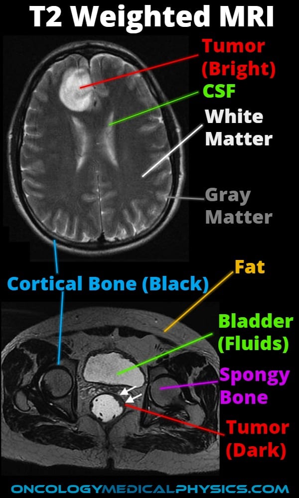
T2 contrast offers higher soft tissue contrast than proton density weighting or T1 weighting. For this reason, T2 weighting is the most widely used weighting scheme in radiation therapy planning. T2 weighting is achieved using a long TR (>2000ms) which allows near full T1 recovery while long TE times (=T2) creates contrast. These long TR times and low signal intensity per excitation make a T2 weighted scan slow compared with T1. The most common T2 weighted sequence is Fast Spin Echo.
Key Point: Cancerous tissue tends to have longer T2 times making tumors appear bright on T2 weighted images, especially when surrounded by edema. This is not always true, see image for examples of bright and dark tumors, but is a good rule of thumb.
Proton Density Weighting
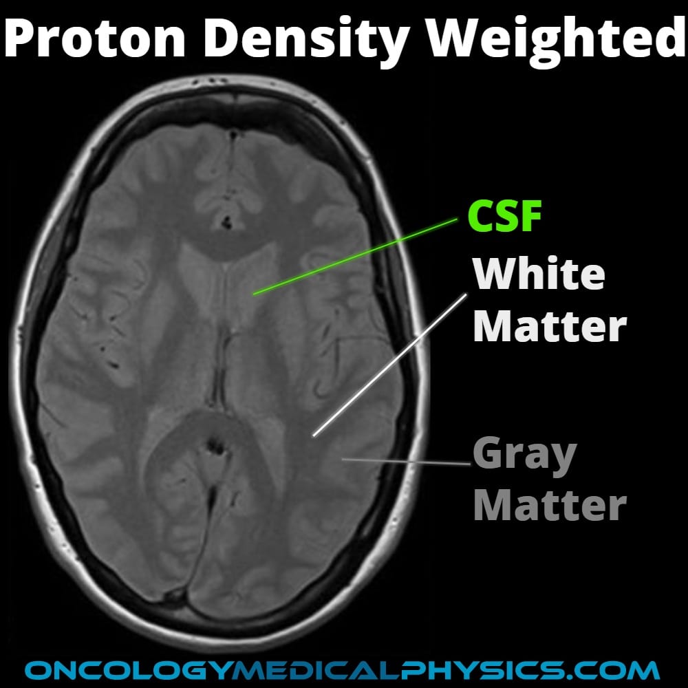
Contrast in a proton density weighted MRI is derived from differences in the number of protons within a voxel. Because most soft tissue has similar proton density, proton density weighting is a low-contrast imaging technique but finds use in evaluation of menisci and brain structures. Proton density weighting is achieved using long TR times (>2000ms) to allow full T1 recovery and very short TE times (< T2) to minimize T2 contrast.

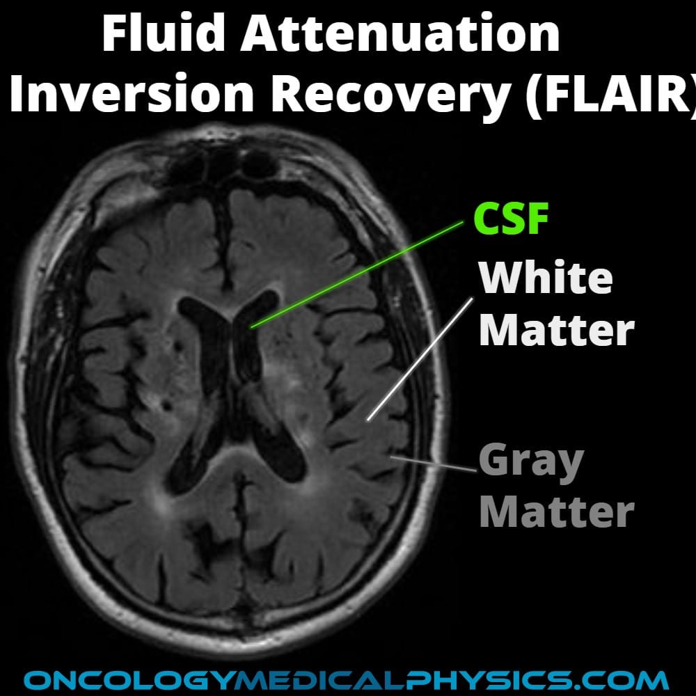
Fluid Attenuated Inversion Recovery (FLAIR)
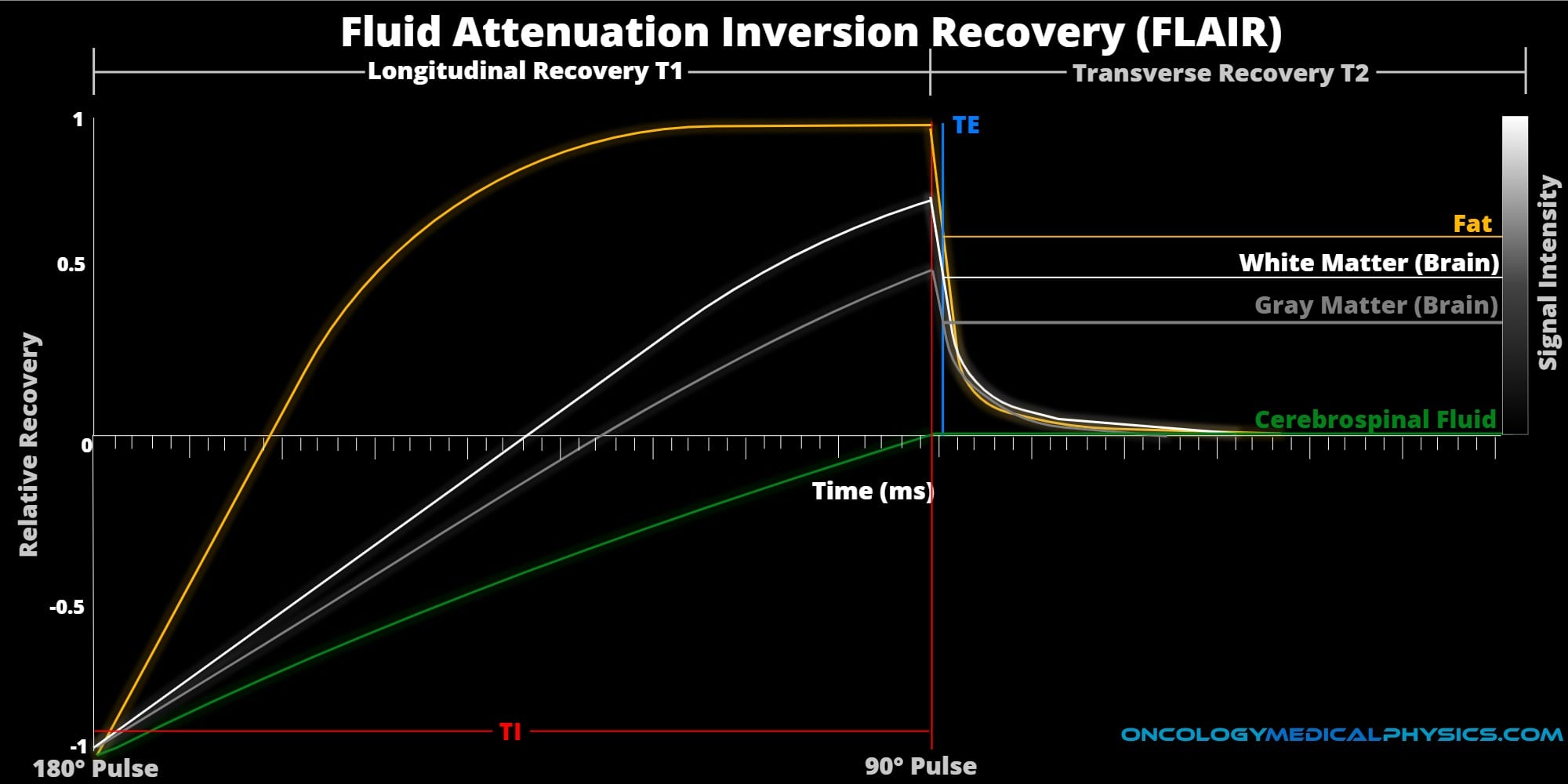
Like all inversion recovery sequences, FLAIR begins with a 180-degree RF pulse to invert the magnetic moment of the effected hydrogen. The 90-degree excitation pulse is timed to coincide with fluid T1 recovery crossing 0 magnetic moment thereby suppressing its signal. FLAIR is most commonly used to suppress cerebrospinal fluid (CSF) in brain scans which can obscure structural information of T2 weighted brain scans. FLAIR finds significant use in distinguishing cerebral edema. This is because cerebral edema appears bright in both T2 weighted and FLAIR scans but FLAIR suppresses CSF, making identification easier.
Diffusion Weighting
Diffusion weighted images register Bownian motion of individual water molecules. This is extremely useful in detecting necrotic regions of a tumor as necrosis appears brighter than the surrounding tissue. Diffusion weighted images may be referred to as an Apparent Diffusion Coefficient (ADC) map.
Contrast Agents
Paramagnetic Iron Oxide
Clinically referred to as SPIO (Super Paramagnetic Iron Oxide) or USPIO (Ultra Small Paramagnetic Iron Oxide), Paramagnetic Iron Oxide primarily impacts T2 times and produces a signal drop in T2* weighted images. A normal liver will absorb SPIO but a metastasized liver will not. This will cause the normal liver to appear dark and the metastasized liver to appear bright.
Navigation
Not a Premium Member?
Sign up today to get access to hundreds of ABR style practice questions.


