Nuclear Tomographic Imaging
PET and SPECT
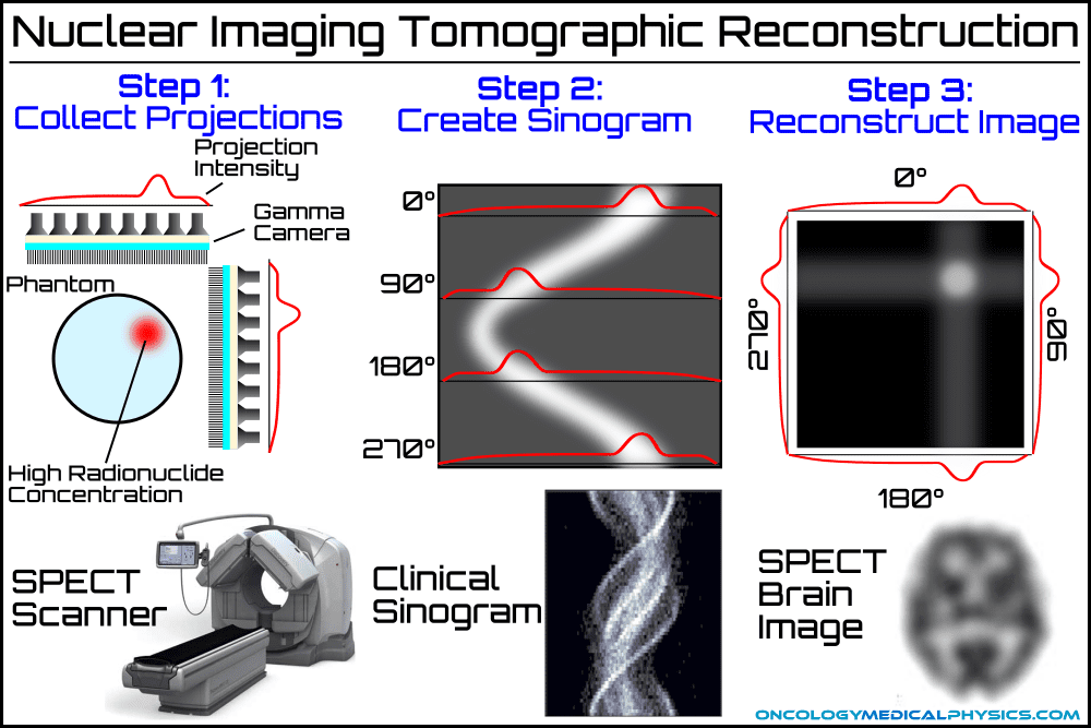
Both Single Photon Emission Computed Tomography (SPECT) and Positron Emission Tomography (PET) involve the creation of 3-dimensional images via computed tomographic reconstruction. The tomographic reconstruction of SPECT and PET images are very similar to CT reconstruction and typically use either filtered back projection or iterative reconstruction. Nuclear tomographic imaging differs from CT imaging in three key areas:
- Nuclear tomographic imaging is used to produce a map of biological activity identified by radiopharmaceutical markers. In contrast, CT imaging produces a map of the linear attenuation coefficient and the density of the body.
- Unlike CT imaging, which uses an external X-ray source, nuclear tomographic imaging relies on radionuclides within the body to generate its information carriers (gamma photons).
- In nuclear imaging, the photon source is within the body. As a result, photon attenuation depends strongly on the source’s path length to the detector. This requires an attenuation correction to prevent sources deep within the patient from appearing darker than superficial sources.
| SPECT | PET | |
|---|---|---|
| Radiation Detectors | Planar Anger Camera | Full 360 degree scintillators |
| Scintillator | NaI(Tl) | LSO, GSO, LYSO |
| Most Common Nuclide | Tc-99m | F-18 (FDG) |
| Attenuation Correction | Patient thickness Attenuation scans CT | CT |
| Photon Vector Determination | Collimator | Coincident photon detection |
| Spatial Resolution | ~10mm FWHM Dependent on orbit and collimator choice | 4-5mm |
Single Photon Emission Computed Tomography (SPECT)
A modern SPECT system consists of 1 to 3 gamma cameras with the dual image design being the most common. The gamma cameras are mounted on a complex gantry system allowing them to image from multiple angles around the patient.
SPECT Imaging Parameters
- Image array
- 64x64 or 128x128
- Collimation
- Parallel collimators are the most common
- Fan beam collimators (a cross between a converging collimator and a pinhole collimator) are used to provide magnification in brain imaging scans
- Data collection modes
- Step-and-shoot collection – data is collected at discrete angles (typically 64-128 positions)
- Continuous collection – data is collected continuously during imager rotation
- Orbit
- Circular – the simplest orbit with the lowest collection efficiency and signal-to-noise ratio
- Elliptical/Contoured – the imager path closely follows the patient’s body. This orbit requires a complex gantry, but provides the highest collection efficiency and signal-to-noise ratio
- Degrees of rotation
- 360° - most common
- 180° - commonly used in cardiac imaging to improve temporal resolution
- Typical imaging dose: 1-5 mSv
Attenuation Correction
Photon attenuation must be determined to avoid a cupping artifact, in which deep seated objects appear darker than superficial objects. Three techniques are commonly used to correct for attenuation:
Thickness Based Estimates
Attenuation may simply be estimated and corrected for based on a standard model and a measure of the patient’s “thickness”.
Attenuation Scans
Attenuation scans are generated using the SPECT/PET scanner’s imager, but using an external photon source. Attenuation scans are able to generate maps of the linear attenuation coefficient called attenuation maps which may be used to correct more accurately for attenuation.
CT Imaging
CT imaging is performed in addition to nuclear tomographic imaging, as it provides both anatomical information and a direct map of attenuation.
Common SPECT Nuclides
Technetium-99m
The most commonly used nuclear imaging radionuclide. Uses include:
-
- Bone
- Renal imaging and function
- Cardiac function
- Breast cancer imaging
- Cerebral perfusion
- Hepatic function
Gamma energy: 140.5 keV
Half-life: 6.007 hours
Production: Tc-99m's short half-life makes remote production and transportation impractical. Instead its parent nuclide, Mo-99, is produced and transported to hospitals as Tc-99m generators. These generators, colloquially known as Moly Cows, produce Tc-99m via beta decay with a half-life of 66 hours. Mo-99 is itself produced in nuclear reactors.
Positron Emission Tomography (PET)
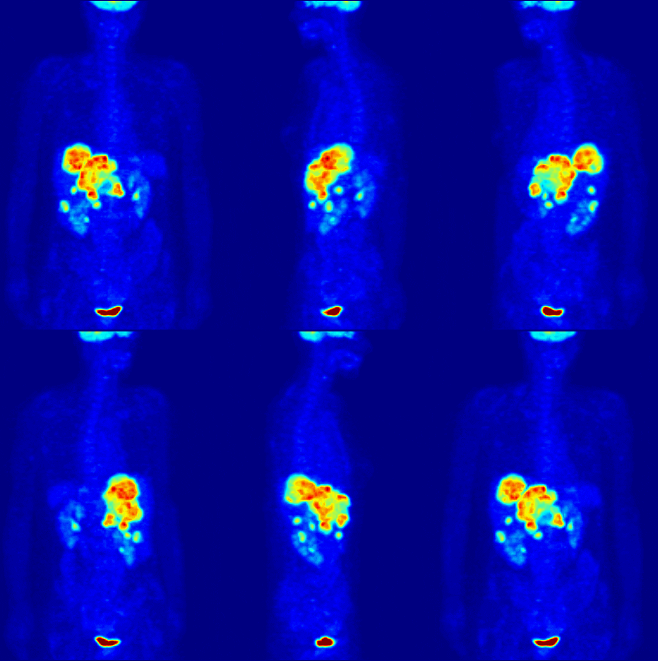
Like SPECT and CT, positron emission tomography (PET) generates 3-dimensional images via tomographic reconstruction of photon projection data. PET imaging is unique in that the photons collected are generated by positron-electron annihilation processes which produce two 511 keV photons which project away from the annihilation site at approximately opposite angles. This unique photon arrangement (two photons traveling in opposite directions) has the unique ability of localizing the site of annihilation to a single point along a path by analyzing the location and timing of photon detection events. PET imaging has experienced great success owing largely to its ability to image metabolic activity via Fluorine-18 fluorodeoxyglucose (FDG).
Design
PET systems are comprised of several rings of gantry-mounted detectors which surround the patient during imaging. The use of multiple detector rings allows for the acquisition of many (over 100) transverse slices simultaneously. The design also improves system sensitivity by being able to detect coincident photon events which are oriented slightly off-axis (nearly, but not quite orthogonal to the bore axis).
Scintillation Crystal
Common NaI(Tl) scintillators used in gamma cameras and SPECT imaging have a low detection efficiency for 511 keV photons generated in PET imaging. As a result, several other scintillators are used; including Lu2SiO2 (LSO), Gd2SiO4O (GSO), and LuxY2-xSiO4O (LYSO). These crystals have higher densities requiring smaller crystals and less self-attenuation of scintillation light. This means that these specialty crystals have higher collection efficiency and superior spatial resolution for PET applications.
Coincidence Detection
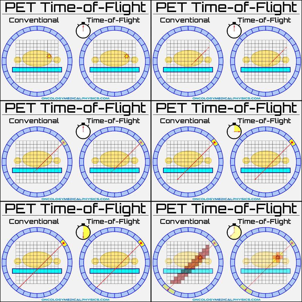
PET scanners can identify multiple detection events occurring at the same time in different areas. These coincident events are assumed to result from a single positron annihilation emitting two photons in opposite directions. Therefore, the source can be assumed to lie on the line connecting the coincident detections. This means that PET scanners do no need collimators to determine the path of incident photons, which greatly increases their collection efficiency.
While coincidence detection removes the need for a physical collimator, it does introduce two new forms of noise:
- Scatter coincidence occurs when one of the annihilation photons is scattered prior to detection
- Random coincidence occurs when two photons arising from separate annihilations are detected simultaneously
Time of Flight
Annihilations photons travel at the speed of light from their point of origin to the detector. The time required for this journey, called the time of flight, is predominately dependent upon the distance traveled. This allows advanced PET scanners to determine the approximate point of origin along the photon’s path by computing the time between coincident events.
Key point: If time-of-flight calculations could perfectly determine the point of annihilation along the photons path, tomographic reconstruction would not be necessary to produce 3D images. Uncertainty in scintillation decay time, along with the speed of light, limits position determination to a few cm and necessitates tomographic reconstruction.
Attenuation Correction
All modern PET scanners are coupled with a CT scanner in a PET/CT system. In a PET/CT system, the patient passes through both the PET and CT bores in a single scan. This CT image allows for precise correction of photon attenuation within the patient.
Limitations on Spatial Resolution
PET scanner spatial resolution is fundamentally limited to a few millimeters for several reasons:
- Positrons travel a small, but non-zero, distance between emission and annihilation. For F-18, this distance is approximately 2.4 mm. This means that annihilations take place outside of tissues which absorb PET radiopharmaceutical markers.
- The 511 keV annihilation photons do not travel in exactly opposing directions. This introduces uncertainty in the exact path that the annihilation photons traveled along.
- Time of flight calculations are dependent upon the time required for the scintillator to emit scintillation photons after an interaction. Although PET scintillation crystals are very prompt (quick to emit photons), scintillation is a decay phenomenon which has some fundamental uncertainties. This dependence limits the determination of the point of origin to within a few centimeters for LSO and LYSO detectors.
Common PET Nuclides
Fluorine-18
Uses: F-18 is used as the marker in fluorine-18 fluorodeoxyglucose (FDG). FDG is a glucose analogue which is absorbed in tissues with high rates of metabolic activity. This makes FDG ideal for imaging many forms of cancer, in differentiating viable tissue from scar tissue in the myocardium, and several other applications.
Half-life: 109.8 minutes
Emission Energy: Positron - 0.633MeV (approximate range of 2.4mm in tissue)
Production: Cyclotron bombardment of O-18 via 18O (p, n) 18F reaction
Typical dose: 7mSv for adult scan (370MBq administered)
Navigation
Not a Premium Member?
Sign up today to get access to hundreds of ABR style practice questions.


