
CT Dosimetry
CT Dose in the United States
Use of computed tomography is increasing and with this increase has come a growing awareness of the population risks involved. As of 2015 as many as 80 million CTs were taken in the U.S. alone. Between 1980 and 2006, CT dose per capita rose 566% from 0.53mSv to 3.0mSv. While the risk of radiation induced cancer is low on an individual level (about 5% per Sv for adults and about 15% per Sv for children) this level of radiation exposure is likely responsible for a significant number of cancer inductions across the U.S. One study (external link) has estimated that CT alone is responsible for as much as 1.5% to 2% of U.S. cancers at the 2007 use level.


Factors Impacting CT Dose
- kVp (Dose ∝ kVp2)
- mAs (Dose ∝ mAs)
- Pitch (Dose ∝ 1/pitch)
- Beam quality
- Patient size and anatomy
- Filtration (bow tie filter shape and material)
- Source to detector distance
- Use of automatic exposure control (AEC)
Measuring CT Dose
CT Dose Phantoms
Two 150mm long PMMA phantoms are used to measure Computed Tomography Dose Index (CTDI). Both have ion chamber drillings along the center and edges for measurement of CTDIw.
- Adult body phantom: 32 cm diameter
- Adult head/pediatric torso phantom: 16cm diameter

Computed Tomography Dose Index (CTDI)
CTDI is defined as the integral of the dose profile along the z-axis of a phantom divided by the nominal beam width.
![]()
- N is the number of slices in a scan
- T is the slice width
CTDI100
CTDI100 is defined as the cumulative dose at the center of a 100mm axial scan.
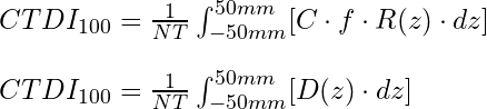
- C is the ion chamber calibration factor (typically ~1.0)
- f is the exposure-to-dose conversion factor (f = 0.87 cGy/R)
- R is the chamber reading
CTDIW
CTDIw is a weighted average of CTDI100 values measured at the center of the phantom and at the edge of the phantom. The Concept of CTDIw was created to give a better sense of integral dose throughout the phantom. Because of attenuation, CTDI may vary by a factor of 2 or more between measurements made at the surface and those made at the center of the phantom.
Typical CTDIw values
- ~8cGy for pediatric/head phantom
- ~3cGy for body phantom
![]()
CTDIVol
CTDIvol normalizes dose from a helical scan with an arbitrary pitch to a pitch of 1 or axial scan. For an axial scan, CTDIvol = CTDIw.
![]()
- I is the table motion per rotation. Unit: mm/rotation
Dose Length Product (DLP)
Practical methods of CTDI measurement integrate dose of 100mm but in a typical scan, much more of the patient will be exposed than 100mm. DLP accounts for differences in scan length.
![]()
Effective Dose
Effective dose is used to estimate the radiation risk from various radiological exams. Effective dose incorporates a weighting factor k which signifies risk to an organ relative to whole body exposure.
![]()
Limitation of Effective Dose
The value of k is dependent upon age but specifies risk to a "standard human" not any individual patient. Further, acrylic cylindrical phantoms are standard for measurement of CTDI. This geometry and difference in material composition means that effective dose is computed on dose data which is fundamentally unrepresentative of a human dose distribution.
Source: AAPM report No. 96
| Under 1 year old | 1 year old | 5 years old | 10 years old | Adult | |
|---|---|---|---|---|---|
| Head and Neck | 0.013 | 0.0085 | 0.0057 | 0.0042 | 0.0031 |
| Head | 0.011 | 0.0067 | 0.0040 | 0.0032 | 0.0021 |
| Neck | 0.017 | 0.012 | 0.011 | 0.0079 | 0.0059 |
| Chest | 0.039 | 0.026 | 0.018 | 0.013 | 0.014 |
| Abdomen & Pelvis | 0.049 | 0.030 | 0.020 | 0.015 | 0.015 |
| Trunk | 0.044 | 0.028 | 0.019 | 0.014 | 0.015 |
Size Specific Dose Estimate
SSDE attempts to make CT dose estimates more applicable to individual patients by applying a conversion factor, fsize, to CTDIvol. The conversion factor, fsize, is found via a lookup table in TG-204 (external link) and is based on the concept of effective diameter (equation below). Effective diameter may also be estimated based on patient age using additional tables with TG-204 (external link). With this modification, SSDE is believed to be accurate to +/-20%.
One interesting aspect of SSDE is that it is found to be only weakly dependent on kVp. Within the range of 80-140kVp, SSDE varies by less than +/-5%.
![]()
![]()
Further Reading
AAPM TG-204 Size-Specific Dose Estimates (SSDE) in Pediatric and Adult Body CT Examinations (External Link)
Key Point: On a per-patient basis, our knowledge of dose is limited to only about +/-20% using SSDE.

Key Point: SSDE should not be used with k-factors to determine effective dose.
Navigation

Not a Member?
Sign up today to get access to hundreds of ABR style practice questions.